Home › Biological sample › Cancer › Liver
Liver cancer biological samples
For Translational Research
The development of drugs and diagnostic tests for the treatment and detection of liver cancer requires conducting studies on biological samples obtained from patients with liver cancer.
A brief overview of the various types of liver cancer and how the services offered by Labtoo contribute to accelerating research and development projects in the pharmaceutical industry.

Biospecimen from liver cancer patients
Tissues from liver cancer patients
Fresh tissues
Frozen tissues (OCT and FF)
FFPE Tissues
Adjacent Healthy Tissues
Blood derivatives from liver cancer patients
-
Plasma
-
PBMC
-
Whole Blood
-
Serum
Biofluids from liver cancer patients
-
Bile
-
Urine
-
Ascitic fluid
-
Other
Associated clinical data from liver cancer patients
-
Age
-
Gender
-
TNM Classification
-
Undergone Treatment
-
Medical Imaging
-
HIV/HBV/HCV status
-
Mutations
-
Other Data (upon request)
Send your request to our team:
What are liver tumors?
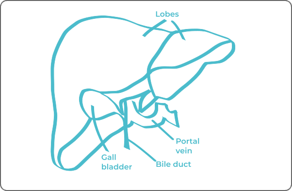
The liver, as a key organ in the digestive system, assumes several crucial functions in the body. It plays an active role in the metabolism of nutrients, regulation of hormonal levels, and detoxification by filtering harmful substances. These processes significantly contribute to maintaining homeostasis, thereby ensuring overall physiological balance.
The major primary cancers of the liver are distinguished into two types, differentiated by their originating cells.
Hepatocellular carcinoma (HCC)
Constituting approximately 85% of liver cancers, arises from the transformation of hepatocytes, the main liver cells.
Risk factors associated with HCC include infections with hepatitis B and C, cirrhosis, and alcoholism.
Various variants of HCC, such as fibrolamellar hepatocellular carcinoma, scirrhous, pleomorphic, or sarcomatoid carcinoma, are identified based on their specific morphology.
Cholangiocarcinoma
Less common, originates from the epithelial cells of the bile ducts, known as cholangiocytes.
| Type of Liver Cancer | Originating Cells | Frequency |
|---|---|---|
| Hepatocellular Carcinoma (HCC) | Hepatocytes | 85% |
| Fibrolamellar HCC | Hepatocytes | Rare |
| Scirrhous HCC | Hepatocytes | Rare |
| Pleomorphic HCC | Hepatocytes | Rare |
| Sarcomatoid HCC | Hepatocytes | Rare |
| Cholangiocarcinoma | Cholangiocytes | 10% |

Due to its significant vascularization, the liver is frequently the site of metastases. Colorectal, breast, lung, gastric, and other cancers can metastasize to the liver. The distinction between primary and metastatic cancer is crucial due to its impact on therapeutic choices and prognosis.
Explore Labtoo's Service for Your Biological Sample Research
Labtoo assists you in sourcing biological samples from liver cancer patients. Our team manages the entire project of transferring biological materials from inception to sample delivery.
- Feasibility assessment of sample availability or clinical collection from referenced clinical centers
- Validation of regulatory aspects
- Establishment of a contractual framework
- Dispatch of desired samples under appropriate conditions
- Transfer of associated clinical data
- Additional analytical and experimental services
The stages and grades of liver cancer
The stage and grade of cancer are commonly used together to provide a comprehensive evaluation of the disease and guide optimal treatment.
The determination of the cancer stage relies primarily on the TNM classification, which assesses the size of the tumor (T), the involvement of lymph nodes by cancer cells (N), and the presence of metastases in other parts of the body (M).
Concurrently, the grade provides an indication of the degree of differentiation of cancer cells. Noted from 1 to 3, Grade G1 indicates well-differentiated cells resembling normal cells, Grade G2 represents moderately differentiated cells, and Grade G3 indicates poorly differentiated cells, suggesting faster and potentially more aggressive growth.
For liver cancer, the Barcelona Clinic Liver Cancer (BCLC) classification is commonly used, defining 4 stages A, B, C, D:
Stage 0
Observation of a tumor 2 cm or less, causing no symptoms, and not invading major blood vessels of the liver.
Stage A
Observation of up to 3 tumors, each less than 3 cm, no symptoms, and no invasion of major blood vessels of the liver.
Stage B
Observation of more than 3 tumors in the liver or 1 to 3 tumors, with at least one larger than 3 cm, no symptoms, and no invasion of major blood vessels of the liver.
Stage C
Invasion of major blood vessels of the liver or spread outside the liver to other parts of the body, causing symptoms.

Rare forms of liver cancer
Occasionally, rare forms of liver cancer develop, among which are:
-
Hepatic Angiosarcoma
This cancer type develops from endothelial cells lining the blood vessels of the liver.
-
Hepatoblastoma
It arises from embryonic cells of the liver and predominantly affects children.
-
Hepatic Neuroendocrine Carcinoma
A highly uncommon case, the tumor originates from neuroendocrine cells of the liver.
Liver cancer treatments and advances
The treatment of liver cancer depends on various parameters, particularly the stage of the disease. For cancers diagnosed at stage A, surgery is typically preferred, involving partial resection of the affected liver or potentially liver transplantation. Chemotherapy is often considered in conjunction with surgery to prevent relapses.
In cases where surgery is not possible or insufficient, different therapeutic alternatives may be considered, such as radiotherapy, cryoablation, and percutaneous radiofrequency tumor destruction.
For cancers classified at stage B, chemoembolization approaches can be used to obstruct the tumor's blood supply.
Immunotherapy has also demonstrated its efficacy in treating advanced liver cancer. It is frequently integrated into targeted therapy strategies, in combination with anti-tumor agents such as bevacizumab or sorafenib.


