Home › Biological sample › Cancer › Lung
Lung cancer biological samples
For Translational Research
The development of drugs and diagnostic tests for the treatment and detection of lung cancer requires conducting studies on various biospecimens collected from breast cancer patients.
A brief overview of the various types of Lung cancer and how the services offered by Labtoo contribute to accelerating research and development projects in the pharmaceutical industry.

Biospecimen from lung cancer patients
Tissues from lung cancer patients
Fresh tissues
Frozen tissues (OCT and FF)
FFPE Tissues
Adjacent Healthy Tissues
Blood derivatives from lung cancer patients
-
Plasma
-
PBMC
-
Whole Blood
-
Serum
Biofluids from lung cancer patients
-
Bronchoalveolar fluid
-
Urine
-
Other
Associated clinical data from lung cancer patients
-
Age
-
Gender
-
TNM Classification
-
Undergone Treatment
-
Medical Imaging
-
HIV/HBV/HCV status
-
Mutations
-
Other Data (upon request)
Send your request to our team:
What are lung tumors ?
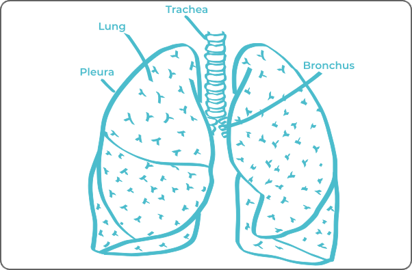
The lungs play a vital role in facilitating gas exchange, promoting blood oxygenation, and eliminating carbon dioxide, thus ensuring the overall vitality of the organism.
Primary lung cancers originate from the epithelial cells of the lungs and are classified into two distinct categories based on the microscopic morphology of tumor cells: Non-small cell lung cancer (NSCLC) and small cell lung cancer (SCLC).
Non-small cell lung cancers (NSCLC)
This is the most common group of tumors.
Including pulmonary adenocarcinoma, squamous cell carcinoma arising from the bronchial epithelial cells, and large cell carcinoma derived from larger epithelial cells in various pulmonary areas.
Small cell lung cancers (SCLC)
They are less common but more invasive, typically originating from neuroendocrine cells in the central part of the lungs.
Predominant types include small cell carcinomas and mixed small cell carcinomas.
| Type of Cancer | Origin Cell | Frequency |
|---|---|---|
| Squamous Cell Carcinoma | Epithelial Cells | ≈ 25 - 30% |
| Adenocarcinoma | Glandular Cells | ≈ 40 - 50% |
| Small Cell Carcinoma | Neuroendocrine Cells | ≈ 15 - 20% |
| Large Cell Carcinoma | Epithelial Cells | ≈ 10 - 15% |
| Carcinoid Tumors | Neuroendocrine Cells | > 5% |
| Pleural Mesothelioma | Mesothelium | Rare |

Lung cancers can also be metastatic, emanating from a malignant tumor in another organ. The distinction between primary and secondary cancer is crucial for determining appropriate management.
Explore Labtoo's Service for Your Biological Sample Research
Labtoo assists you in sourcing biological samples from lung cancer patients. Our team manages the entire project of transferring biological materials from inception to sample delivery.
- Feasibility assessment of sample availability or clinical collection from referenced clinical centers
- Validation of regulatory aspects
- Establishment of a contractual framework
- Dispatch of desired samples under appropriate conditions
- Transfer of associated clinical data
- Additional analytical and experimental services
The stages and grades of lung cancer
The stage and grade of cancer are commonly used together to provide a comprehensive assessment of the disease and guide optimal treatment.
The determination of cancer stage primarily relies on the TNM classification, which evaluates the tumor size (T), involvement of lymph nodes by cancer cells (N), and the presence of metastases in other parts of the body (M). Concurrently, the grade provides an indication of the degree of differentiation of cancer cells.
Regarding grades, denoted from 1 to 3, Grade G1 indicates well-differentiated cells resembling normal cells, Grade G2 represents moderately differentiated cells, and Grade G3 indicates poorly differentiated cells, suggesting faster and potentially more aggressive growth.
For lung cancer, the stages are defined as follows:
Stage 0
Cancer cells remain localized in the lining of the airways or alveolar sacs.
Stage I A
The lung tumor has a maximum size of 3 cm.
Stage I B
The tumor, measuring between 3 cm and 4 cm, may invade different areas such as the airway and visceral pleura, potentially causing inflammation of lung tissues.
Stage II A
The tumor, measuring between 4 cm and 5 cm, may invade different areas, including the airway and visceral pleura, potentially causing inflammation of lung tissues.
Stage II B
The tumor measures 5 cm and has spread to lymph nodes near the bronchi
Or the tumor measures between 5 and 7 cm
Or it has invaded the parietal pleura, thoracic wall, and phrenic nerve.
Or, there are at least two tumors in the same lobe.
Stage III A
The tumor measures 5 cm and has spread to lymph nodes of the trachea or near the bronchi or both.
Or, the cancer is more than 5 cm and has invaded a nearby body part (diaphragm, heart, trachea, etc.).
Or, the tumor is more than 5 cm, and there are at least 2 tumors in the same lung.
Stage III B
The tumor measures 5 cm, and cancer has spread to lymph nodes on the opposite side of the trachea or lung, or to lymph nodes in the lower part of the neck.
Or, the tumor measures more than 5 cm, and cancer has also spread to lymph nodes next to the trachea, or to lymph nodes beneath the region where the trachea divides into the bronchus.
Stage III C
The tumor measures more than 5 cm, or there is more than one tumor in a different lobe of the same lung.
Spread of cancer to lymph nodes on the opposite side of the trachea or lung, or to lymph nodes located in the lower part of the neck.
Stade IV
Cancer has spread to other parts of the body (metastases).

Rare forms of lung cancer
Occasionally, rare forms of lung cancer develop, among which are:
-
Lung Neuroendocrine Tumors (NETs)
Represent a type of cancer arising from neuroendocrine cells present in lung tissues. The two common variants of NETs are typical carcinoid tumors and atypical carcinoid tumors.
-
Pulmonary Apex Tumor
Also known as Pancoast tumor, it may manifest different histological types, such as squamous cell carcinoma or adenocarcinoma. Its designation comes from its location at the rounded top of the lung, a feature that increases the likelihood of infiltration into the chest wall.
-
Pleural Mesothelioma
This is a type of cancer that develops in the cells of the pleural mesothelium, the membrane surrounding the lungs. This disease is often associated with asbestos exposure, and its origin lies in the malignant transformation of mesothelial cells.
Lung cancer treatments and advances
The treatment of lung cancer depends on the type of cancer, its stage, and the patient's physical condition. The main therapeutic modalities include surgery, radiotherapy, chemotherapy, immunotherapy, and targeted therapies.
-
Surgery: Surgery is often considered for non-small cell lung cancers. It may involve resection of part of the lung (lobectomy), an entire lobe (pneumonectomy), or a smaller part of lung tissue (segmentectomy or wedge resection).
-
Radiotherapy: Radiotherapy uses radiation to destroy cancer cells or prevent their growth. It may be administered before surgery to shrink the tumor, after surgery to eliminate residual cancer cells, or as the primary treatment for patients who cannot undergo surgery.
-
Chemotherapy: Chemotherapy uses cytotoxic drugs to target and kill cancer cells. It can be administered before or after surgery or as the main treatment to control tumor growth.
-
Immunotherapy: Immunotherapy stimulates the immune system to recognize and fight cancer cells. It is increasingly used in lung cancer treatment, especially for non-small cell forms.
-
Targeted Therapies: These drugs target specific abnormalities present in cancer cells. They are used when specific genetic mutations are identified. Targeted therapies are often used for non-small cell lung cancers.


