Home › Biological sample › Cancer › Skin
Skin cancer biological samples
For research applications
The development of drugs and diagnostic tests for the treatment and detection of skin cancer requires conducting studies on biological samples obtained from patients with skin cancer.
A brief overview of the various skin cancer types and how the services offered by Labtoo contribute to accelerating research and development projects in the pharmaceutical industry.

Biospecimen from skin cancer patients
Tissues from skin cancer patients
Fresh tissues
Frozen tissues (OCT and FF)
FFPE Tissues
Adjacent Healthy Tissues
Blood derivatives from skin cancer patients
-
Plasma
-
PBMC
-
Whole Blood
-
Serum
Biofluids from skin cancer patients
-
Interstitial fluid
-
Other
Associated clinical data from skin cancer patients
-
Age
-
Gender
-
TNM Classification
-
Undergone Treatment
-
Medical Imaging
-
HIV/HBV/HCV status
-
Mutations
-
Other Data (upon request)
Send your request to our team:
What are skin tumors?
The most common skin cancers are distinguished into three types based on the type of cells in which they originate: The basal cell carcinoma, squamous cell carcinoma, as well as melanoma.
Basal cell carcinoma
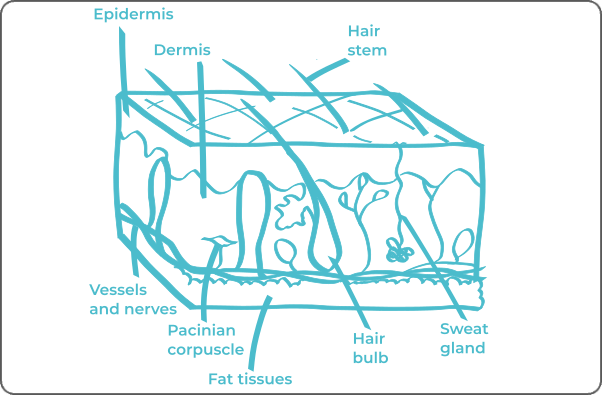
This is the most common skin cancer, developing from keratinocytes, the deepest cells of the epidermis.
Although it can appear anywhere on the body, it is more common on the head and neck.
This type of carcinoma poses a low risk of metastasis.
Squamous cell carcinoma
Also known as spinocellular carcinoma, less common, it forms from epithelial cells in the intermediate layer of the epidermis.
It can appear on any part of the body, but its preferential areas are those exposed to the sun.
The risk of metastasis is higher with this type of carcinoma.
Melanoma
Although less common, melanoma is the most serious of skin cancers.
It develops from melanocytes, responsible for skin color.
This type of cancer carries an increased risk of metastasis.
|
Type of Skin Cancer
|
Origin Cell
|
Frequency (Relative)
|
|---|---|---|
|
Basal Cell Carcinoma
|
Basal Cells
|
70%
|
|
Squamous Cell Carcinoma
|
Squamous Cells
|
20%
|
|
Melanoma
|
Melanocytes
|
10%
|
|
Kaposi's Sarcoma
|
Endothelial Cells
|
>1%
|
|
Cutaneous Lymphoma
|
Lymphatic Cells
|
1-5%
|
|
Merkel Cell Cancer
|
Merkel Cells
|
>1%
|
|
Dermatofibrosarcoma
|
Soft Tissues of the Skin
|
>1%
|
|
Annexal Carcinoma
|
Annexal Glands (Sweat and Sebaceous)
|
>1%
|

Skin cancers can be metastatic. Since the dermis is highly vascularized, tumors may spread through lymphatic or blood vessels to infect other organs.
Explore Labtoo's Service for Your Biological Sample Research
Labtoo assists you in sourcing biological samples from skin cancer patients. Our team manages the entire project of transferring biological materials from inception to sample delivery.
- Feasibility assessment of sample availability or clinical collection from referenced clinical centers
- Validation of regulatory aspects
- Establishment of a contractual framework
- Dispatch of desired samples under appropriate conditions
- Transfer of associated clinical data
- Additional analytical and experimental services
The stages and grades of skin cancer
The stage and grade of cancer are commonly used together to provide a comprehensive assessment of the disease and guide optimal treatment.
The determination of cancer stage primarily relies on the TNM classification, which evaluates the tumor size (T), involvement of lymph nodes by cancer cells (N), and the presence of metastases in other parts of the body (M). Concurrently, the grade provides an indication of the degree of differentiation of cancer cells.
Regarding grades, denoted from 1 to 3, Grade G1 indicates well-differentiated cells resembling normal cells, Grade G2 represents moderately differentiated cells, and Grade G3 indicates poorly differentiated cells, suggesting faster and potentially more aggressive growth.
For skin cancer, the stages are defined as follows:
Carcinoma
Stage 0
Cancer cells are limited to the epidermis.
Stage I
Tumor size up to 2 cm.
Stage II
Tumor size between 2 and 4 cm.
Stage III
At least one statement
- Tumor larger than 4 cm
- Mild spread to a neighboring bone.
- Invasion of nerves or deeper development than the epidermis.
- Spread to a lymph node with a maximum size of 3 cm.
Stage IV A
At least one statement
- Extension to a lymph node larger than 3 cm.
- Spread to multiple lymph nodes measuring less than 6 cm.
- Extension to a lymph node larger than 6 cm.
Stage IV B
Spread to other parts of the body (metastases).
Melanoma
Stage 0
Cancer cells are limited to the epidermis.
Stage I A
Tumor thickness of 0.8 mm or less, without ulceration,
OR thickness between 0.8 mm and 1 mm with possible ulceration.
Stage I B
Tumor thickness between 1 mm and 2 mm, without ulceration.
Stage II A
At least one statement
- Tumor thickness between 1 mm and 2 mm with ulceration.
- Tumor thickness between 2 mm and 4 mm, without ulceration.
Stage II B
At least one statement
- Tumor thickness between 2 mm and 4 mm with ulceration.
- Tumor thickness over 4 mm without ulceration.
Stage III
Spread to at least 1 lymph node.
Stage IV
Spread to other parts of the body (metastases).

Rare forms of skin cancer
Occasionally, rare forms of liver cancer develop, among which are:
-
Merkel Cell Carcinomas
Formed from neuroendocrine cells in the upper layer of the skin.
-
Dermatofibrosarcoma Protuberans
Or Darier-Ferrand disease. There emerges in the deep layers of the skin, involving fibroblasts, the cells responsible for the production of skin collagen.
-
Adnexal Carcinomas
Group of skin cancers forming from adnexal structures such as sebaceous glands, sweat glands, hair follicles, and nails. This includes sebaceous, apocrine, and eccrine carcinomas.
-
Cutaneous Lymphoma
A type of cancer affecting lymphatic cells present in the skin.
-
Kaposi's Sarcoma
A type of skin cancer developing in the endothelial cells lining blood and lymphatic vessels. Often associated with Human Herpesvirus 8 (HHV-8).
Skin cancer treatments and advances
The treatment of skin cancers varies depending on the stage of the disease, the patient's age, and the type of tumor.
Surgery remains the primary treatment in all cases. Depending on the tumor's postoperative response or if surgery is not feasible, various complementary methods may be considered.
These approaches include chemotherapy, immunotherapy, radiotherapy, cryotherapy, photodynamic therapy, as well as targeted therapies specifically aiming at precise proteins present in the tumor.
The treatment choice is personalized based on a comprehensive assessment of the patient's clinical situation by the medical team.


