Home › Biological sample › Cancer › Uterine
Uterine cancer biological samples
For Translational Research
The development of drugs and diagnostic tests for the treatment and detection of uterine cancer requires conducting studies on biological samples obtained from patients with uterine cancer.
A brief overview of the various types of uterine cancer and how the services offered by Labtoo contribute to accelerating research and development projects in the pharmaceutical industry.

Biospecimen from uterine cancer patients
Tissues from uterine cancer patients
Fresh tissues
Frozen tissues (OCT and FF)
FFPE Tissues
Adjacent Healthy Tissues
Blood derivatives from uterine cancer patients
-
Plasma
-
PBMC
-
Whole Blood
-
Serum
Biofluids from uterine cancer patients
-
Endometrial lavage
-
Vaginal swab
-
Urine
-
Other
Associated clinical data from uterine cancer patients
-
Age
-
Gender
-
TNM Classification
-
Undergone Treatment
-
Medical Imaging
-
HIV/HBV/HCV status
-
Mutations
-
Other Data (upon request)
Send your request to our team:
What are uterine tumors?
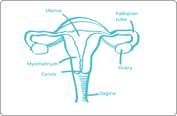
The uterus, a central organ of the female reproductive system, plays a crucial role in embryonic development by ensuring the encapsulation and nutrition of the fetus until its maturation.
Regarding uterine cancers, they can be categorized into two distinct types based on their location: cervical cancer and endometrial cancer.
Cervical cancer
It primarily encompasses two major histological types, squamous cell carcinoma and adenocarcinoma.
Squamous cell carcinoma can be keratinizing or non-keratinizing, originating from the squamous cells of the external surface of the cervix.
Adenocarcinoma arises from the glandular cells of the cervix, with subtypes such as mucinous adenocarcinoma and endometrioid adenocarcinoma. Some cases may exhibit characteristics of both histological types, forming an adenosquamous tumor.
Approximately 90% of cervical cancer cases are triggered by human papillomavirus (HPV) infection.
Endometrial cancer
Also known as endometrial carcinoma or uterine body cancer, it differs in origin and is not associated with viral infection.
It primarily originates from the epithelial cells of the uterine mucosa.
| Cancer Type | Tumor Name | Cellular Origin | Frequency |
| Cervical Cancer | Squamous Cell Carcinoma | Squamous epithelial cells | ≈ 70 - 90% cervical cancers |
| Adénocarcinoma | Glandular cells | ≈ 10 - 25% of cervical cancer | |
| Adenosquamous Tumor | Squamous and glandular cells | Rare | |
| Endometrial Cancer | Endometrial Carcinoma | Epithelial cells of the uterine mucosa | ≈ 80% of endometrial cancer cases |
| Carcinosarcoma | Epithelial and mesenchymal cells | Rare | |

Explore Labtoo's Service for Your Biological Sample Research
Labtoo assists you in sourcing biological samples from uterine cancer patients. Our team manages the entire project of transferring biological materials from inception to sample delivery.
- Feasibility assessment of sample availability or clinical collection from referenced clinical centers
- Validation of regulatory aspects
- Establishment of a contractual framework
- Dispatch of desired samples under appropriate conditions
- Transfer of associated clinical data
- Additional analytical and experimental services
The stages and grades of uterine cancer
The stage and grade of cancer are commonly used together to provide a comprehensive assessment of the disease and guide optimal treatment.
The determination of the cancer stage relies primarily on the TNM classification, which evaluates the tumor size (T), involvement of lymph nodes by cancer cells (N), and the presence of metastases in other parts of the body (M).
In parallel, the grade provides an index of the degree of differentiation of cancer cells, graded from 1 to 3. Grade G1 indicates well-differentiated cells resembling normal cells, Grade G2 represents moderately differentiated cells, and Grade G3 indicates poorly differentiated cells, suggesting faster and potentially more aggressive growth.
Stages of cervical cancer
Stage I A
Lesion measuring a maximum of 3 mm in depth and 7 mm in width
Stage I A2
Lesion measuring between 3 and 5 mm in depth and a maximum of 7 mm in width
Stage I B1
Lesion measuring less than 2 cm in the major axis
Stage I B2
Lesion measuring between 2 and 4 cm in the major axis
Stage II A1
Lesion measuring less than 2cm in the major axis
Stage II A2
Lesion measuring more than 4 cm in the major axis
Stage III A
The lesion has extended to the lower third of the vagina without affecting the pelvic wall.
Stage III B
The lesion has invaded the pelvic wall or obstructs a ureter, causing hydronephrosis (kidney swelling), or impairs the proper functioning of a kidney.
Stage IV A
The lesion has spread to the bladder, rectum, or beyond the pelvis.
Stage IV B
Cancer cells have migrated to other parts of the body.
Stages of endometrial cancer
Stage I A
The tumor is in the endometrium, invasion is less than half of the myometrium.
Stage I B
Invasion of half of the myometrium.
Stage II
Invasion of the cervix.
Stage III A
Invasion of the uterine serosa and/or ovaries, fallopian tubes, and ligaments.
Stage III B
Extension to the vagina and/or parametria
Stage III C
Invasion of pelvic and/or para-aortic lymph nodes.
Stage IV A
Invasion of the bladder or intestine lining.
Stage IV B
Presence of metastasis

Rare forms of uterine cancer
Occasionally, rare forms of uterine cancer develop, among which are:
-
Choriocarcinoma
A rare tumor occurring in the uterus during pregnancy.
-
Uterine muscle sarcoma
Also known as uterine leiomyosarcoma (LMS), a tumor taking shape from the smooth muscle cells of the uterus.
-
Carcinosarcoma
A rare tumor composed of sarcomatous and carcinomatous elements.
Uterine cancer treatments and advances
The treatment of uterine cancer requires an individualized, multidisciplinary approach.
-
In the case of cervical cancer, treatment modalities are based primarily on surgery, radiotherapy, and chemotherapy. These interventions can be applied in isolation or in combination, depending on the type of tumor and its stage of development. Targeted therapy, which specifically targets tumor molecules, is also used. The predominant targeted therapy strategy is to hinder tumor angiogenesis by neutralizing the protein responsible for vascular growth, often using monoclonal antibodies such as bevacizumab.
-
For the cancer of the corpus uteri, the choice of treatment depends on the stage of the disease. Surgery is generally preferred, especially for tumors up to stage 2. Radiotherapy may be used after surgery in certain cases, to prevent recurrence. Chemotherapy is used less frequently and is mainly reserved for high-grade carcinomas, using agents such as cisplatin or carboplatin combined with paclitaxel. In the case of stage 3 cancer, when surgery or radiotherapy is not feasible, hormone therapy may be considered, often combined with the use of megestrol and medroxyprogesterone.
The use of immunotherapy in cases of uterine cancer is promising, although currently in the experimental stage.


