Home › Biological sample › Cancer › Testicular
Testicular cancer biological samples
For research applications
The development of drugs and diagnostic tests for the treatment and detection of testicular cancer requires conducting studies on biological samples obtained from patients with testicular cancer.
A brief overview of the various types of testicular cancer and how the services offered by Labtoo contribute to accelerating research and development projects in the pharmaceutical industry.

Biospecimen from testicular cancer patients
Tissues from testicular cancer patients (tumoral & normal adjacent)
Fresh resection & biopsy
Frozen samples (OCT & FF)
FFPE blocks & slides
Blood derivatives from testicular cancer patients
-
Whole Blood
-
PBMC
-
Plasma
-
Serum
Biofluids from testicular cancer patients
-
Semen
-
Urine
-
Other
Associated clinical data from testicular cancer patients
-
Age
-
Gender
-
TNM Classification
-
Undergone Treatment
-
Medical Imaging
-
HIV/HBV/HCV status
-
Mutations
-
Other Data (upon request)
Send your request to our team:
What are testicular tumors?
Testicular cancer, constituting less than 5% of male cancers, is generally unilateral. Unlike ovarian tumors, dominated by epithelial forms, the predominant testicular tumors are derived from germ cells.
Germ cell tumors are classified into two main categories:
Seminomatous tumors
These emerge from degenerated spermatogonial stem cells, primarily affecting men aged 35 to 45.
Non-seminomatous tumors
Arising from the degeneration of stem cells capable of differentiating into various cell types, they affect younger men, between 16 and 35.
This group includes several histological types, such as embryonal carcinomas, teratomas, and choriocarcinomas, which differ in morphology and degree of cellular differentiation.
| Type of Cancer | Tumor Name | Cell of Origin | Frequency |
| Seminomatous Tumors | Seminomatous Tumor | Germ Cells | ≈ 60% |
| Non-Seminomatous Tumors (35-40%) | Embryonal Carcinoma | Stem Cells | >5% |
| Teratoma | Stem Cells | ≈ 5 - 10% | |
| Choriocarcinoma | Stem Cells | Rare | |
| Non-Germ Cell Tumors | Non-Germ Cell Tumor | Various Cells | ≈ 5% |

It should be noted that, although rarer, non-germ cell cancers of the testicles exist. Examples include rete testis adenocarcinoma and malignant mesothelioma of the tunica vaginalis.
Testicular cancer can metastasize, with the abdominal lymph nodes being frequently affected, sometimes extending to the lungs or liver. It is noteworthy that it is exceptional for a secondary tumor from another organ to migrate to the testicles.
Explore Labtoo's Service for Your Biological Sample Research
Labtoo assists you in sourcing biological samples from testicular cancer patients. Our team manages the entire project of transferring biological materials from inception to sample delivery.
- Feasibility assessment of sample availability or clinical collection from referenced clinical centers
- Validation of regulatory aspects
- Establishment of a contractual framework
- Dispatch of desired samples under appropriate conditions
- Transfer of associated clinical data
- Additional analytical and experimental services
The stages and grades of testicular cancer
The stage and grade of cancer are commonly used together to provide a comprehensive assessment of the disease and guide optimal treatment.
The determination of cancer stage primarily relies on the TNM classification, which evaluates the tumor size (T), involvement of lymph nodes by cancer cells (N), and the presence of metastases in other parts of the body (M). Concurrently, the grade provides an indication of the degree of differentiation of cancer cells.
Regarding grades, denoted from 1 to 3, Grade G1 indicates well-differentiated cells resembling normal cells, Grade G2 represents moderately differentiated cells, and Grade G3 indicates poorly differentiated cells, suggesting faster and potentially more aggressive growth.
For testicular cancer, the stages are defined as follows:
Stage 0
Presence of a precancerous condition in the testicle, called germ cell neoplasia in situ or intratubular germ cell neoplasia.
Stage I A
The tumor is in the testicle and epididymis.
Possibility of spread to the inner layer of the testicle membrane (albuginea).
Normal tumor marker levels.
Stage I B
Either the tumor is in the testicle and epididymis, it has spread to the blood vessels or lymphatics of the testicle.
Or it has invaded the tunica vaginalis.
Or it has invaded the spermatic cord or scrotum, with a possibility of spread to blood vessels or lymphatics.
Stage I C
The tumor is in the testicle, spermatic cord, or scrotum.
It has a higher-than-normal tumor marker level.
Stage II A
The tumor has spread to at least one inguinal lymph node.
They measure no more than 2 cm.
Stage II B
The tumor has spread to at least one inguinal lymph node.
They measure between 2 and 5 cm.
Stage II C
The tumor has spread to at least one inguinal lymph node.
They measure more than 5 cm.
Stage III
Cancer has spread to lymph nodes not in the groin or to the lungs, or it has spread to a more distant part of the body.
The cancer becomes metastatic.

Rare forms of testicular cancer
Occasionally, rare forms of testicular cancer develop, among which are:
-
The lymphomas of the testicle
Classified among non-Hodgkin's cancers, constitute another rare variant of non-germinal testicular cancer.
-
Paratesticular rhabdomyosarcoma
This exceptional form of cancer is characterized by an origin in the soft tissues surrounding the testicle.
-
Adenocarcinoma of the rete testis
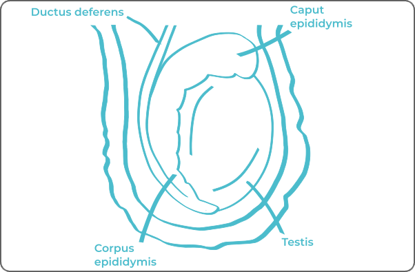
It forms from the glandular (or non-glandular) epithelial cells of the rete testis. The rete testis functions to transport sperm to the epididymis.
-
Malignant mesothelioma of the tunica vaginalis
A rare form of cancer that develops from the layers of cells covering most internal organs of the body, the mesothelium.
Testicular cancer treatments and advances
The treatment of testicular cancer depends on the type of cancer, its stage, and the patient's physical condition. The main therapeutic modalities for addressing this condition include:
-
Surgery: An essential step in treating testicular tumors. The surgeon will perform an orchidectomy, meaning the removal of the testicle carrying the tumor. In rare cases, partial orchidectomy (preserving hormonal and reproductive functions) may be performed.
-
Radiotherapy: Can be applied after surgery. The goal is to administer high-energy rays to the site of tumor development (retroperitoneal lymph nodes) to kill cancer cells. Sometimes used in protocols for seminomatous tumors, but its use is increasingly limited due to side effects.
-
Chemotherapy: Chemotherapy employs cytotoxic drugs to target and kill cancer cells. Often administered after surgery in the case of testicular cancer.
Immunotherapy and targeted therapy are currently not widely used methods in cases of testicular cancers.


